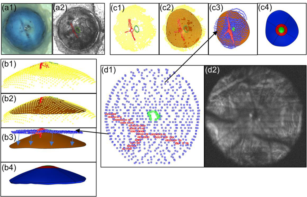
Fig. 3.
Registration of thickness data with DIC data. a1. Image of eye without speckle pattern applied. a2 Eye with speckle pattern applied and LPCAs (red) and optic nerve (green) delineated. Panel c (top view) and panel b (side view) show the same steps from different viewpoints. b1 and c1 Optic nerve (green) and LPCA (red) delineations from DIC image mapped to 3D coordinates using DIC facet map (yellow). b2 and c2 Surface (orange) fit through the DIC facets forming the posterior surface of the sclera model. b3 and c3 SD-OCT delineations of the sclera (blue), LPCAs (red), and optic nerve (green) manually registered to DIC data. Arrows in b3 indicate how the thickness map was projected to the posterior model surface. b4 and c4 Model after thickness data was applied and the model was split up into eight regions. The ONH region is shown in green, the peripapillary regions are shown in red, and the peripheral regions are in blue. d1 SD-OCT delineations of the sclera (blue), LPCAs (red), and optic nerve (green). d2 Average intensity projection of slices from an SD-OCT volume in which the LPCAs and optic nerve are visible

