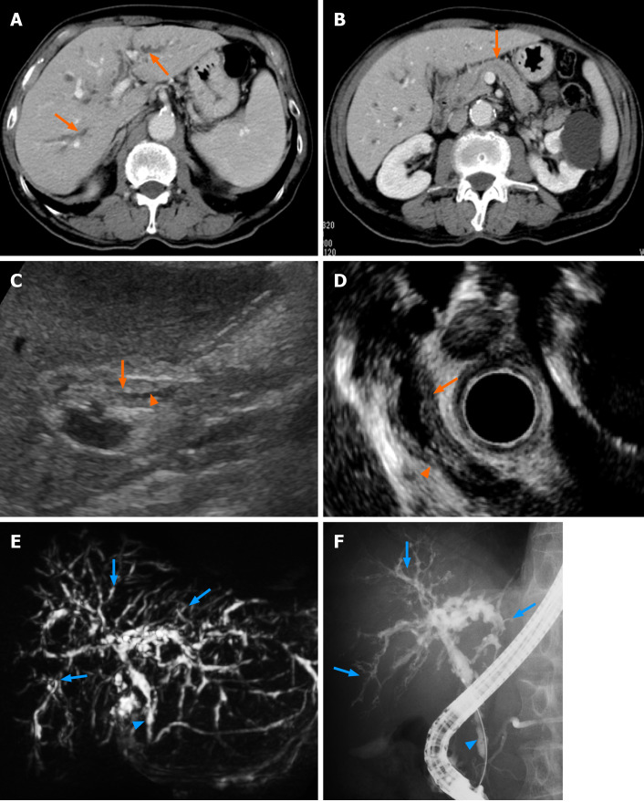
Figure 1.
Contrast-enhanced computed tomography showed stricture and mild dilatation of intrahepatic bile ducts and its wall thickening with an enhance effect (A, orange arrows). No significant swelling of the pancreas and the dilatation of main pancreatic duct were observed (B, orange arrow); C: Abdominal ultrasonography and D: Endoscopic ultrasonography showed thickening of the bile duct wall (C and D, orange arrows) and stenosis of the bile duct (C and D, orange arrowheads) in the lower bile duct. Magnetic resonance cholangiopancreatography and endoscopic retrograde cholangiopancreatography revealed stenosis of the lower bile duct (E and F, blue arrowheads) and intrahepatic bile ducts (E and F, blue arrows).

