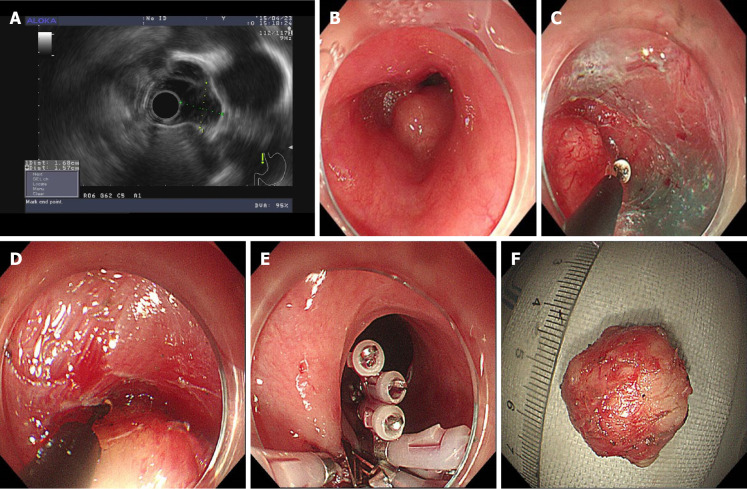
Figure 2.
Case 2 Steps of submucosal tunneling endoscopic resection. A: Endoscopic ultrasound showed the lesion arose from the muscular layer and was misdiagnosed as leiomyoma. The blood flow was not obvious, and the lesion was near the aorta; B: The submucosal tumor located in the middle of the thoracic esophagus; C: A submucosal tunnel was created; D: The tumor was dissected from the muscular layer in the submucosal tunnel; E: Closure of the submucosal tunnel with clips; F: The excised lesion.

