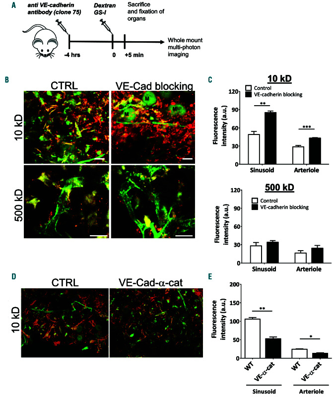
Figure 2.
Anti-VE-cadherin antibodies increase bone marrow vascular permeability in homeostatic conditions and after irradiation. (A) Schematic summary of experimental set up. (B) Bone marrow (BM) imaging after injection of fluorescent dyes with in green GS-I for vessel labeling and in red 10 and 500 kDa dextran as indicated. M: megakaryocytes. (C) Quantification of vascular permeability in arterioles and sinusoids in metaphysis and diaphysis after blocking VE-cadherin (10 kD: n=7-8 per group, 500 kD: n=4 per group). (D) BM imaging of wild-type and VE-cadherin-α-catenin chimera mice after injection of fluorescent dyes with in green GSI for vessel labeling and in red 10 kDa dextran (n=3-4 per group). (E) Quantification of vascular permeability in arterioles and sinusoids in wild-type and VE-cadherin- α-catenin chimera mice as indicated. Scale bars: 25 mm, ctrl: control.

