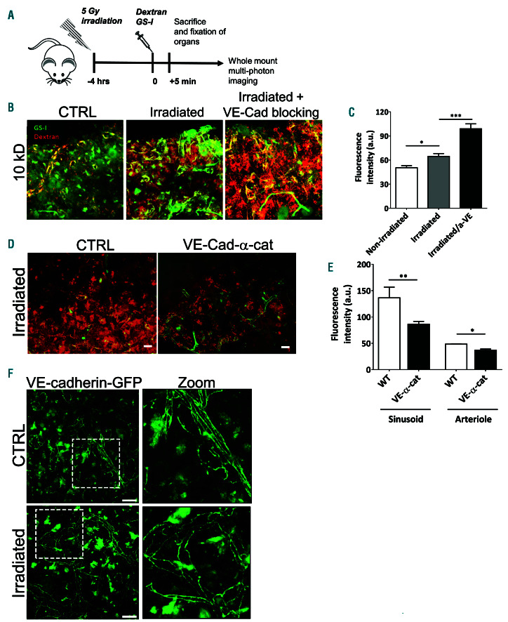
Figure 3.
Reduced bone marrow vascular permeability in VE-cadherin- α-catenin fusion mice. (A) Schematic summary of experimental set up. (B) Bone marrow (BM) imaging of wild-type, VE-cadherin-α-catenin chimera mice and supplemented with anti-VE-cadherin antibody after injection of fluorescent dyes with in green GS-I for vessel labeling and in red 10 kDa dextran (n=3-4 per group). (C) Quantification of vascular permeability in sinusoids in wild-type, VE-cadherin-α-catenin chimera mice and supplemented with anti-VE-cad antibody as indicated. (D) BM imaging of wild-type and VE-cadherin-α-catenin chimera mice after 5 Gy irradiation after injection of fluorescent dyes with in green GS-I for vessel labeling and in red 10 kDa dextran (n=3-4 per group). (E) Quantification of vascular permeability in ar terioles and sinusoids in wild-type and VE-cadherin-α-catenin chimera mice upon irradiation as indicated. (F) BM vasculature using a VE-cadherin-GFP knock-in mice showed proper VE-cadherin lining in control and upon irradiation conditions. Images on right are zoom from dotted box. Scale bars: 25 mm; ctrl: control.

