Figure 7.
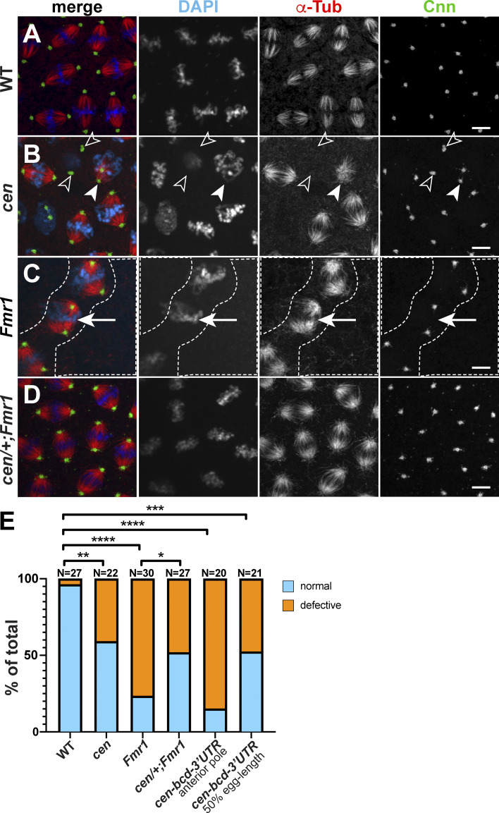
Cen and FMRP ensure proper mitosis. Maximum-intensity projections of metaphase NC 11 embryos from the indicated genotypes stained for β-Tub to label microtubules (red), Cnn (green), and DAPI (blue). (A) WT show uniform bipolar mitotic spindles. (B) cen embryo with reduced microtubules (open arrowheads) and incomplete centrosome separation (closed arrowheads). (C) Fmr1 embryo with nuclear fallout (dashed lines) and bent spindles (arrows). (D) Hemizygosity for cen partially rescues Fmr1 mutants. (E) Frequency of spindle defects from n = 27 WT, 22 cen, 30 Fmr1, 27 cen/+;Fmr1, 20 cen-bcd-3′UTR (anterior), and 21 cen-bcd-3′UTR (50% egg length) embryos. *, P < 0.05; **, P < 0.01; ***, P < 0.001; ****, P < 0.0001 by Fisher’s exact test. Scale bars: 5 µm.

