Figure S2.
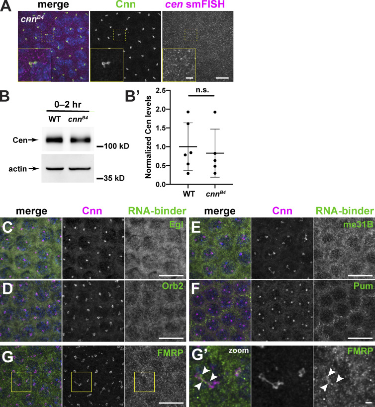
cen mRNA granule formation requires the centrosome scaffold. (A) Image shows immunofluorescence for Cnn (green) and cen smFISH (magenta) in an NC 12 cnnB4 embryo. Boxed region is enlarged in inset. Note the absence of large pericentrosomal cen mRNA granules. (B) Immunoblots show Cen protein content in 0–2-h (up to NC 14) WT and cnnB4 lysates. Actin is used as a loading control. (B′) Each dot represents the levels of Cen normalized to the mean relative expression of the actin load control. n.s., not significant (P = 0.672) by unpaired t test from n = 3 independent biological replicates, with n = 2 technical replicates run on the same gel. (C–G′) Images show interphase NC 12 embryos stained for Cnn (magenta) and antibodies for the indicated RNA-binding proteins (RNA-binder, green): Egl (C), Orb2 (D), me31B (E), Pum (F), and FMRP (G). (G′) Inset from G; arrowheads, FMRP overlapping with Cnn. Scale bars: 10 µm; 2 µm (insets).

