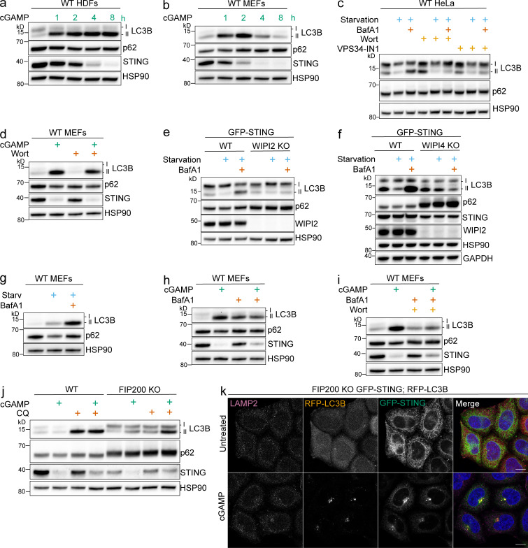
Figure S1.
Experiments supplementary to Figs. 1 and 2. (a and b) Primary HDFs (a) and primary MEFs (b) were treated with 60 μg/ml cGAMP, and cell lysates were collected at the indicated time points. (c) WT HeLa cells were incubated in starvation media (HBSS with Ca2+ and Mg2+) alone and with 100 nM BafA1 and with or without wortmannin (200 nM) or VPS34-IN1 (300 nM) for 4 h. All the following experiments were treated the same unless specified. (d) MEFs were treated with 60 μg/ml cGAMP alone and with wortmannin for 2 h. All the following experiments using MEFs use the same conditions. (e and f) WT, WIPI2 KO (e), and WIPI 4KO (f) HeLa cells stably expressing GFP-STING were incubated in starvation media alone and with BafA1 for 4 h. (g) MEFs were incubated in starvation media alone and with BafA1 for 4 h. (h and i) MEFs were incubated with cGAMP alone and with BafA1 (h) or with BafA1 and wortmannin (i). (j) WT and FIP200 KO HeLa cells were incubated with 15 µg/ml cGAMP alone and with 20 μM chloroquine (CQ) for 8 h. (k) Representative Airyscan-processed confocal imaging of FIP200 KO HeLa cells with stable expression of RFP-LC3B and GFP-STING treated with cGAMP (60 µg/ml) for 8 h, fixed, and immunostained for endogenous LAMP2. Scale bar, 10 µm. Western blotting experiments were independently replicated two (b, c, e, and g) or three (a, d, f, h, i, and j) times. Starv, starvation; Wort, wortmannin.

