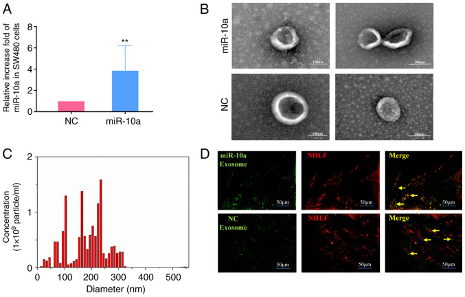
Figure 3.
Identification and tracer analysis of exosomes from SW480 cells. (A) Expression of miR-10a in SW480 cells transfected with mimics-miR-10a (miR-10a) and NC; the isolated exosomes were detected by reverse transcription-quantitative PCR. U6 snRNA was used as an endogenous control. (B) Exosomes released by different groups of SW480 cells, as indicated, were detected by electron microscopy (left panel: magnification, ×100; right panel: magnification, ×200). (C) NanoSight particle diameter analysis of exosomes released by SW480 cells. (D) Fluorescence imaging of the delivery of DiO-labeled exosomes (green) to DiL-labeled NHLFs (red). Yellow arrows represent delivered exosomes. Representative images are presented (magnification, ×200). The experiments were performed at least in triplicate. **P<0.01 vs. NC. NHLFs, normal human lung fibroblasts; miR-10a, microRNA-10a; NC, negative control.

