Figure 1.
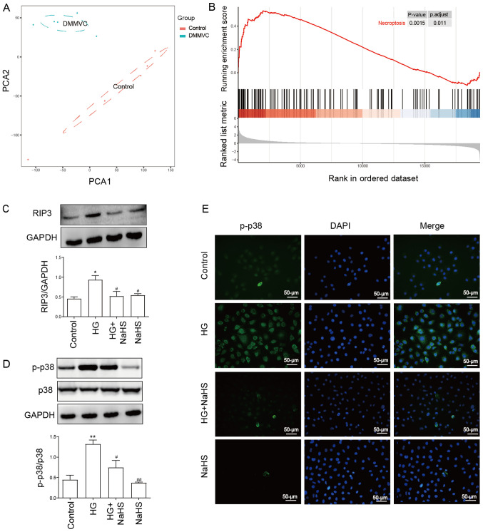
Exogenous NaHS attenuates the HG-induced upregulation of the expression levels of RIP3 and p-p38 in HUVECs. (A) PCA of the GSE43950 dataset obtained from the Gene Expression Omnibus database. Samples including DMMVC and control were separated into two cluster. (B) Gene Set Enrichment Analysis for all genes in the GSE43950 dataset. Genes involved in the necroptosis pathway were enriched in diabetes with microvascular diseases. Expression levels of (C) RIP3 and (D) p-p38/p38 ratio were analyzed and semi-quantified using western blotting. HUVECs were pretreated with or without 400 µM NaHS for 30 min prior to exposure to 40 mM HG. (E) Representative micrographs of immunofluorescence staining of HUVECs with an anti-p-p38 antibody (green) and the fluorescent nuclear stain DAPI (blue) following the indicated treatments. Scale bar, 50 µm. Data are presented as the mean ± SEM (n=3). *P<0.05, **P<0.01 vs. control group; #P<0.05, ##P<0.01, vs. HG group. RIP3, receptor-interacting protein 3; p-, phosphorylated; PCA, principal component analysis; DMMVC, diabetes with microvascular disease; HUVECs, human umbilical vein endothelial cells; NaHS, sodium hydrosulfide; HG, high glucose.

