Fig. 7.
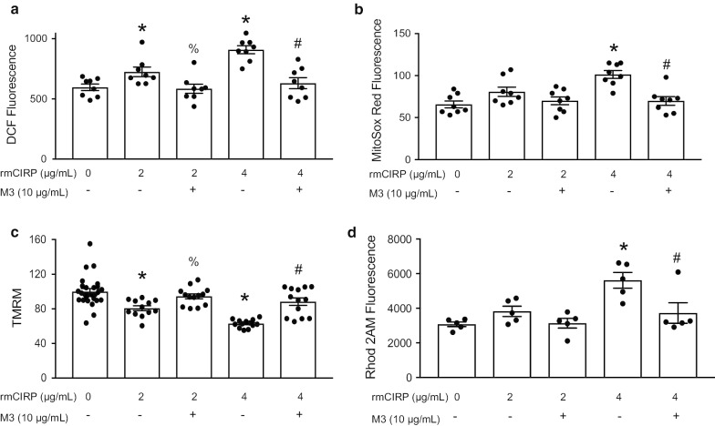
rmCIRP increases oxidative stress in cardiomyocytes and causes mitochondrial dysfunction. Primary murine neonatal cardiomyocytes were isolated. Cardiomyocyte and mitochondrial oxidative stress were quantified using a DCF (N = 8/group) and b MitoSOX Red fluorescence (N = 8/group). Mitochondrial membrane potential and calcium levels was assessed using c Tetramethylrhodamine methyl ester (TMRM) (PBS = 27 and 13 per group for rest) and d Rohd2 AM (N = 5/group) fluorescence. After rmCIRP stimulation, there was a significant dose-dependent increase in cardiomyocyte and mitochondrial-specific reactive oxygen species; this was ameliorated by M3 (a, b). Stimulation of cardiomyocytes with rmCIRP resulted in a reduction in mitochondrial membrane potential while M3 treatment returned membrane potential to near baseline (c). rmCIRP caused mitochondrial calcium overload; this was inhibited by M3 (d). Data shown are the results of two to three independent experiments. Multiple groups were compared by one-way ANOVA and Tukey method (*p < 0.05 vs. PBS control; % p < 0.05 vs 2 µg/mL rmCIRP, #p < 0.05 vs 4 µg/mL rmCIRP)

