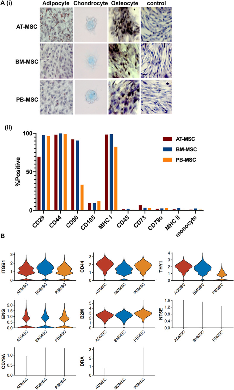
Fig. 1.

Characterization of equine mesenchymal stromal cells (MSCs) isolated from 3 donor-matched tissue sources. a 40x images of adipose tissue (AT-), bone marrow (BM-) and peripheral blood (PB-) derived MSC after in vitro differentiation into adipocytes (oil red O), chondrocytes (alcian blue) and osteocytes (alizarin red). Undifferentiated cells are shown as controls (hematoxylin) (i) and cellular expression patterns of proteins determined by the International Society for Cellular Therapy (ISCT) to be used for MSC immunophenotyping, as detected by flow cytometry (ii). b sc-RNAseq violin plots of the expression levels of the genes corresponding to proteins used for MSC characterization
