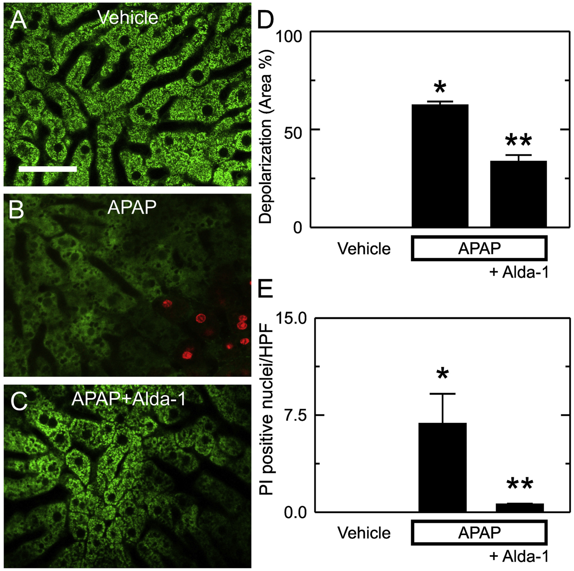
Fig. 6.

Alda-1 decreases mitochondrial depolarization and cell death after APAP overdose. Animals were infused with rhodamine 123 (green fluorescence) and PI (red fluorescence) 6 h after treatment with vehicle only, APAP, and APAP plus Alda-1. Livers were imaged 10 min later using two-photon intravital microscopy. Shown are representative images of Vehicle (A), APAP (B) and APAP plus Alda-1 (C). Bar is 100 μm. For each liver imaged, 10 random images were obtained using a 30× objective lens and quantified for depolarized area (D) and PI-positive nuclei per high power field (E). Values are means ± SEM (n = 3/group). *, p < .05 vs. Vehicle; **, p < .05 vs. APAP.
