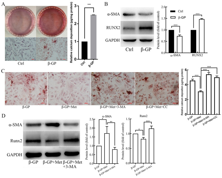
Figure 1.
The effects of β-GP, Met, 3-MA and CC on VSMC calcification. VSMCs were treated with β-GP (10 mmol/l) to induce calcification and with metformin (500 µmol/l) to examine its effect on calcification. 3-MA (5 mmol/l) and CC (10 µmol/l) were added to the cells to investigate the effects of autophagy and the AMPK signaling pathway, respectively. (A) Alizarin red S staining and O-cresolphthalein complexone method calcium quantitation showing calcium deposition in VSMCs treated with vehicle or β-GP. (B) Western blots showing protein levels of α-SMA and Runx2 in VSMCs treated with vehicle or β-GP. (C) Alizarin red S staining and O-cresolphthalein complexone method calcium quantitation showing calcium deposition in VSMCs treated with β-GP, MET, 3-MA, or CC. (D) Western blots showing protein levels of α-SMA and Runx2 in VSMCs treated with β-GP, Met, or 3-MA. Western bands of interest were normalized against GAPDH, and data are provided as relative density ratios compared with control group or β-GP group. Scale bars in Alizarin red S staining represent 100 µm. Representative images are shown. Data are presented as mean ± SEM, n=3. *P<0.05, **P<0.01, ***P<0.001. β-GP, β-glycerophosphate; Met, metformin; 3-MA, 3-methyladenine; CC, compound C; VSMC, vascular smooth muscle cell; α-SMA, α-smooth muscle actin; RUNX2, runt-related transcription factor 2.

