Figure 5.
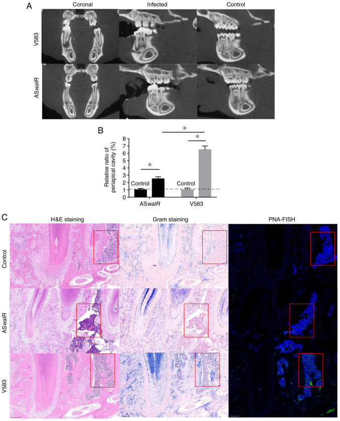
ASwalR overexpression reduces the periapical lesion size and inhibits bacterial aggregation. (A) Reconstructed images for micro-CT scanning of rat periapical lesions. (B) Relative periapical cavity levels (%) were calculated n=10. *P<0.05. (C) Samples were obtained from infected periapical lesions at 4 weeks. HE-stained histological slices (left lane; scale bars, 100 µm) and Gram-stained samples (middle lane; scale bars, 100 µm). The red boxes indicated inflammatory cells in the HE-staining or bacteria in the Gram-staining. The presence of fluorescent Enterococcus faecalis was identified with a PNA-FISH probe for bacterial 16S rRNA (right lane; scale bars, 100 µm). ASwalR, walR antisense RNA; HE, hematoxylin-eosin; PNA-FISH, peptide nucleic acid- fluorescence in situ hybridization.

