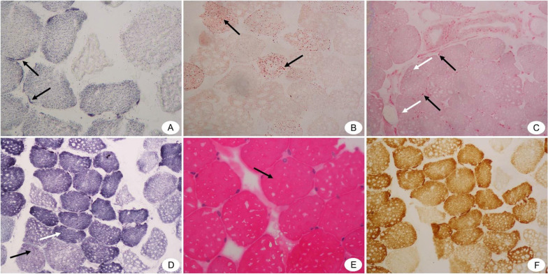
Figure 2.
Pathological results of biopsies from right deltoid muscle of the WD patient (Under light microscope). (A) SDH staining (×400) shows hyperchromatism around type I muscle fibers (shown by black arrows). (B) ORO staining(×200) shows that Lipid droplets in some type I muscle fibers are slightly finer (shown by black arrows). (C) ACP staining(×200) shows that A small number of positive particles are seen in the perimysium (shown by black arrows) and endomysium (shown by white arrows). (D) NADH staining(×200) shows that the muscle fiber structure is a little disordered. Type I muscle fibers are dominant, and there is a small group of group distribution phenomenon (shown by white arrow). The black arrow indicates the type 2 muscle fibers. (E) HE staining(×400)shows that the muscle fibers are blunt round (shown by black arrow). (F) There is no obvious abnormality in COX staining(×200).

