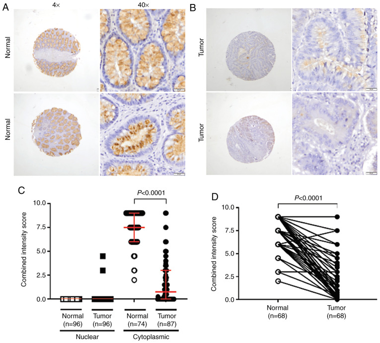
Figure 4.
IHC staining of TFF3. The slides were viewed by light microscopy (×40 magnification). (A) For normal colonic mucosa, TFF3 cytoplasmic staining was mainly associated with the supra- and perinuclear cytoplasm of epithelial cells in both the basal and luminal portions of the colonic crypts. Low to null staining was evident at the nuclear/globular level. (B) TFF3 was lower in cancer cells, and expressed mainly in the supra-nuclear cytoplasm. (C) Scatter plot showing that TFF3 IHC staining is mostly cytoplasmic and lower in tumors, relative to normal mucosa (P<0.0001, Mann-Whitney U test for non-matched data). (D) TFF3 expression was lower in 95.6% of available normal-tumor matching pairs (P<0.0001, Wilcoxon matched-pairs signed rank test). TFF3, trefoil factor 3; IHC, immunohistochemistry.

