Figure 4.
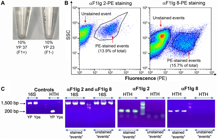
αF1Ig 2 and αF1Ig 8 recognize Y. pestis (YP) in a mixed bacterial community. (A) A mixed bacterial community of Y. pseudotuberculosis, Pseudomonas fluorescens, Escherichia coli, and Bacillus anthracis Sterne (represented equally at 22.5% each) was spiked with YP (10% representation) grown either at 37°C (YP 37, F1+) or 23°C (YP 23, F1–). The resulting cell suspensions were stained with αF1Ig 2–phycoerythrin (PE). After one wash, the bacterial pellet containing YP 37 (F1+) appeared lightly pink, whereas the pellet containing YP 23 (F1–) appeared white. (B) The mixed community spiked with YP 37 (F1+), stained with either αF1Ig 2–PE or αF1Ig 8–PE was analyzed by flow cytometry. Proportionally, 13.9% (αF1Ig 2–PE staining) and 15.7% (αF1Ig 8–PE staining) of the total events were fluorescent (gated events detected by side scatter, SSC, and PE fluorescence). (C) Single cells sorted during the flow experiment depicted in B were lysed. The resulting DNA was used as template for PCR amplification of either the 16S rRNA–encoding region (16S RNA, present in any bacteria, 1,500 bp in length) or a fragment (~200 bp) unique to the genome of Y. pestis (putative “helix-turn-helix” protein [HTH]). While flow-sorted stained and unstained single events tested positive for the 16S rRNA (showing that the detected events were actually bacteria), only the fluorescently labeled bacteria tested positive for HTH and were thus confirmed to be Y. pestis. The discrepancy between the intended YP representation and the representation measured by flow was likely due to inaccuracy in the relationship between cell density (OD600) and cell number, which for the various bacteria used in this experiment was assumed to be OD600 = 0.2 → ×108 cells/mL.

