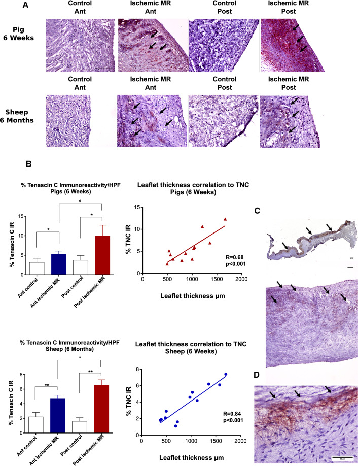
Fig. 3.
Leaflet TNC expression in the ischemic MR animal models and patients’ samples. Tenascin C (TNC) staining (black arrows) of anterior (Ant) and posterior (Post) mitral leaflet. Scale bar 100 µm (a) Quantitative analyses of TNC staining and its correlation to leaflet thickness; *p < 0.01, **p < 0.001 (b). TNC staining (black arrows) was mostly present in the atrial side of the posterior mitral leaflet in the pigs (upper panel) and in the sheep (lower panel); (c) representative TNC staining of mitral leaflet from human patient with ischemic MR (d)

