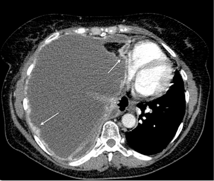
Fig. 11.

Malignant pleural mesothelioma in a 71-year-old man. Axial contrast-enhanced CT image at the level of descending thoracic aorta shows enhancing irregular pleural thickening involves the costal and mediastinal pleural (white arrows). A massive right pleural effusion with contralateral mediastinal shift is also seen.
