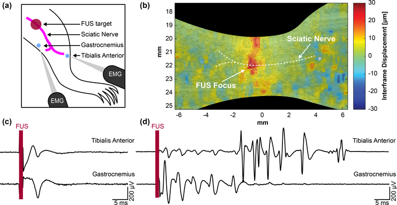
Fig. 4.
(a) Diagram showing mouse leg topology and relative locations of the stimulation and recording sites. (b) Displacement imaging validates focal position onto the sciatic nerve and a region of interest can be taken to acquire average interframe nerve displacement at the focus. A 28 MPa (MI = 6.5), 1 ms pulse was used to map the displacement. (c) Example traces showing single CMAPs from a single FUS stimulus. (d) Example traces of multiple CMAPs from a single FUS stimulus.

