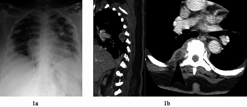
Figure 1. Chest x-ray and CT chest with contrast. 1a: Plain chest x-ray showing decreased lung volume, cardiomegaly, and multiple opacities concerning of coronavirus disease 2019 (COVID-19) infection. 1b: CT chest with contrast demonstrating ring enhancing lesions (arrows) at base of right lung at thoracic vertebrae 5 and 6 in sagittal and axial views.

