Fig. 1.
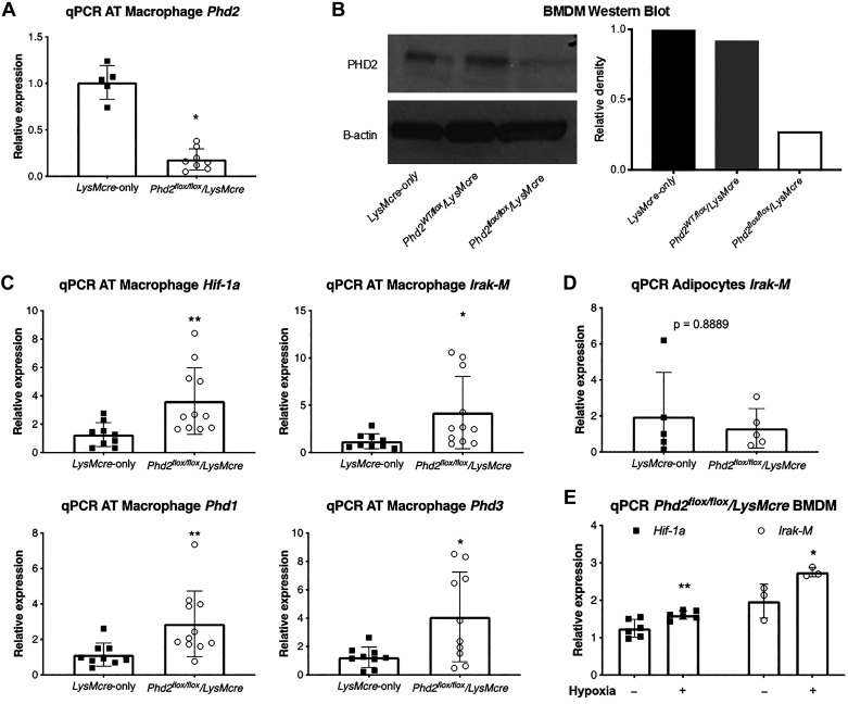
Phd2 inhibition in macrophages leads to overexpression of Hif-1α and its downstream effector Irak-M. A: qPCR analysis of Phd2 expression in macrophages isolated from adipose tissue (AT) of Phd2flox/flox/LysMcre and LysMcre control mice (n = 5–8 per group). B: Western blot analysis of bone marrow-derived macrophages (BMDM) of Phd2flox/flox/LysMcre and control mice (samples pooled from 2 to 3 mice per group). Right: quantitative analysis of band density by ImageJ software. C: qPCR analysis of Hif-1a in adipose tissue macrophages in mice fed with high-fat diet (HFD), together with Hif-1α downstream effector Irak-M, and two other isoforms of Hif-1α prolyl-hydroxylases Phd1 and Phd3 (n = 9–11 per group). D: qPCR analysis of Irak-M in isolated adipocytes of Phd2flox/flox/LysMcre and LysMcre control mice fed a HFD. E: qPCR analysis of BMDM of Phd2flox/flox/LysMcre mice exposed to normoxic or hypoxic gas mixture. The readings are normalized by Hprt and Gapdh. **P < 0.01, *P < 0.05.

