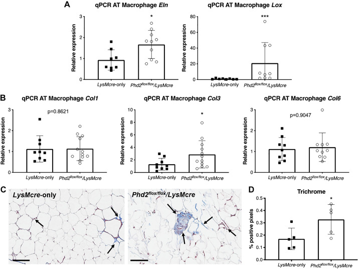
Fig. 3.
Knockdown of Phd2 in macrophages leads to increased adipose tissue fibrosis after 12 wk of HFD. A: qPCR analysis of elastin (Eln) and lysyl oxidase (Lox) in LysMcre-only control and Phd2flox/flox/LysMcre mice (n = 9–10 per group). B: qPCR analysis of collagens I, III, and VI (Col1, Col3, Col6) in LysMcre and Phd2flox/flox/LysMcre mice (n = 9–12 per group). C: Masson’s trichrome stain (arrows) for adipose tissue of LysMcre (left) and Phd2flox/flox/LysMcre transgenic mice (right). Black bars indicate 100 μm. D: quantitative analysis of positive pixels for trichrome stain (n = 5 animals per group, mean of 10 tissue sections per animal). *P < 0.05, ***P < 0.001.

