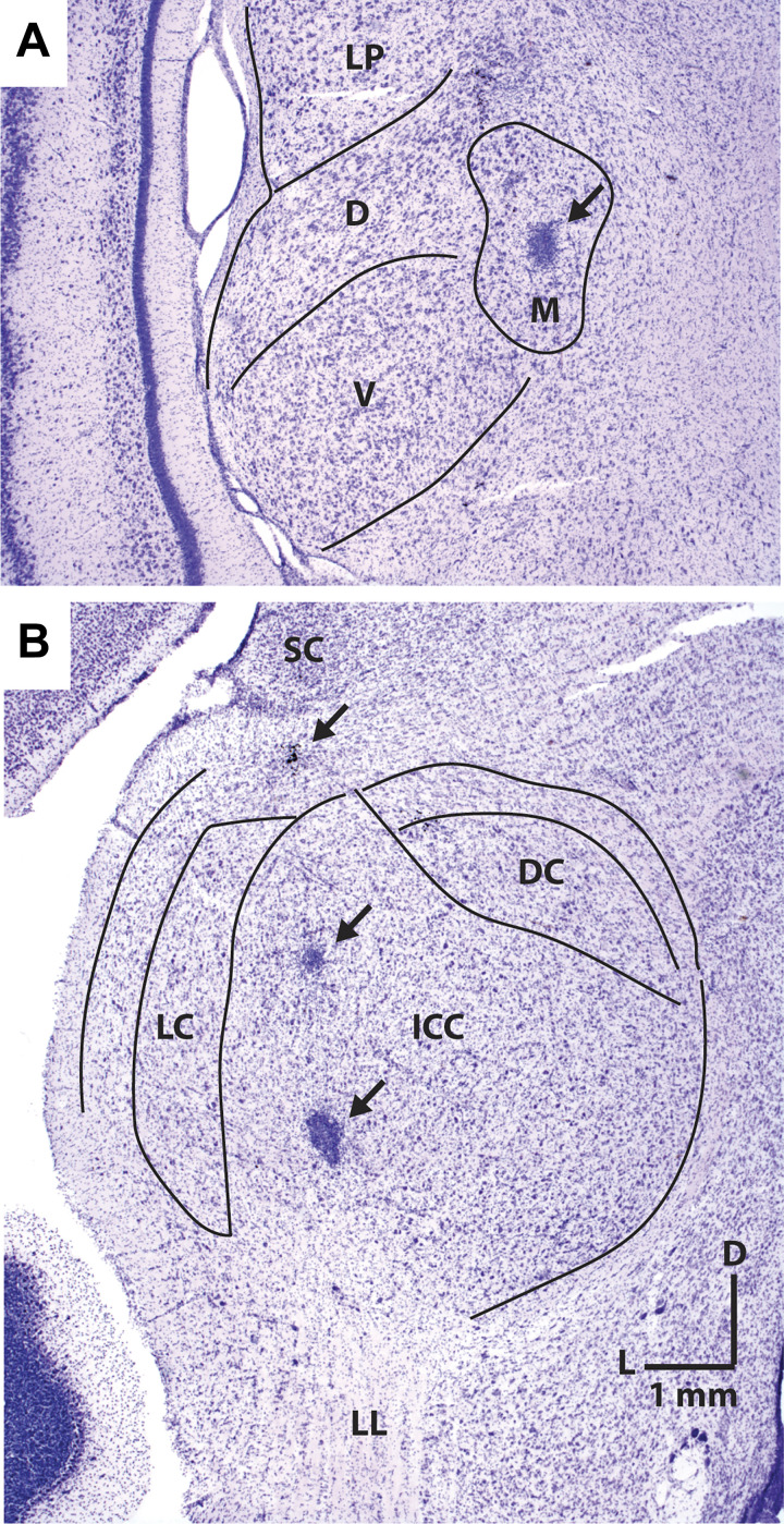
Fig. 2.
Histological sections in transverse planes that show electrolytic lesions of recording sites marked by arrows in the medial geniculate body (A) and the inferior colliculus (B). D, dorsal; DC, dorsal cortex; ICC, central nucleus of inferior colliculus; L, lateral; LC, lateral cortex; LL, lateral lemniscus; LP, lateral posterior; M, medial; SC, superior colliculus; V, ventral.

