Image 4.
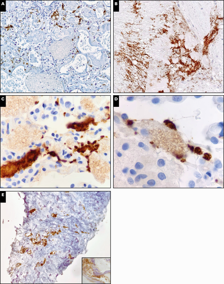
Examples of pathogen-specific staining patterns identified with CD61 immunostaining. A, Discrete staining of capillaries adjacent to yeast colony, with faint surface staining of yeast forms adjacent to platelets (×10). B, Large patch of positive staining likely representing angioinvasion in aspergillosis, with direct platelet-hyphal interactions observed (×20). C, D, Platelet aggregates adjacent to fungal colonies with surface staining of yeasts in pneumocystis infection (×40). E, Staining of vessels at the periphery of invasive zygomycosis and evidence of direct fungal-hyphae interactions (×20; inset, ×40).

