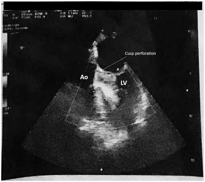
Figure 4.
Monochromatic (black and white) printed colour Doppler images (mid-oesophageal five-chamber view, 0-degrees) showing mild eccentric aortic valve regurgitation by a tiny perforation (arrow) due to the endocarditis vegetation (in the cubicles of the ICU, isolated and specifically dedicated to COVID-19 patients, the echocardiography machine had only a black and white printer). Ao, ascending aorta; LV, left ventricle.

