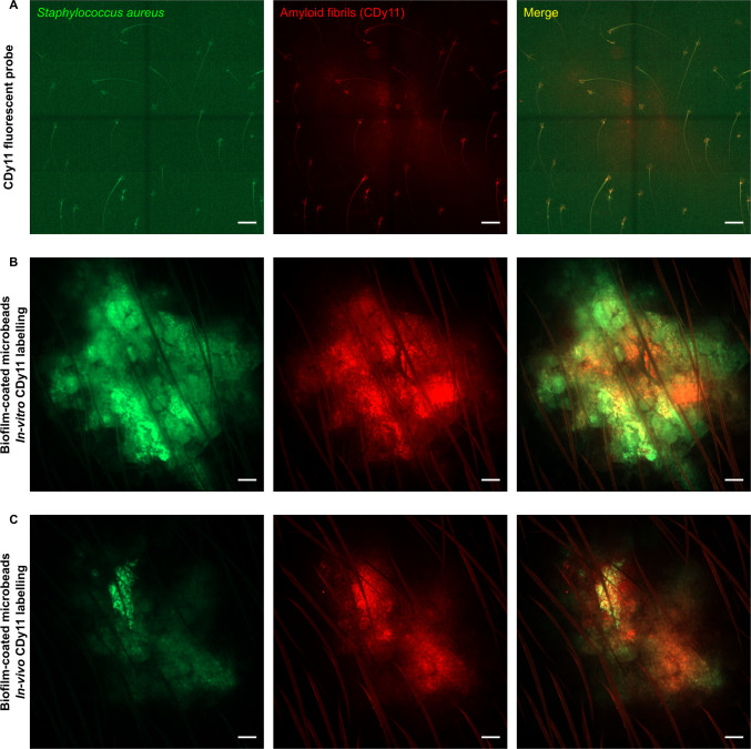
Fig 3. Intravital confocal imaging of CDy11 labelled biofilm-coated microbeads.
Intravital confocal imaging after microinjection of only the CDy11 fluorescent probe (A), of biofilm-coated microbeads after in vitro CDy11 labelling (B) or of biofilm-coated microbeads after in vivo CDy11 labelling (C) in the ear pinna of WT C57BL/6 mice. (A to C) Images show maximum intensity projections of GFP (green) and CDy11 (red) fluorescence. The merged image shows maximum intensity projections for both fluorescence channels. Scale bar: 100 μm.

