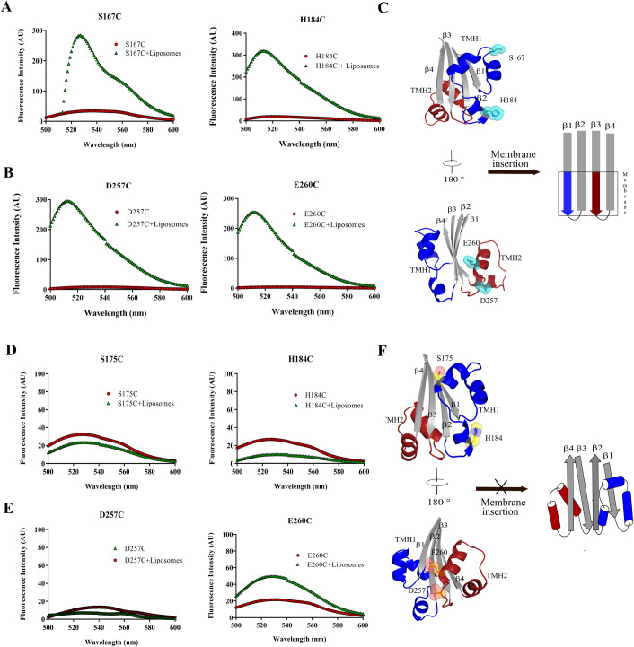
Fig 3. Ply-NH is unable to form transmembrane β-hairpins (TMH1 and TMH2) Fluorescence intensities of all the labelled proteins with and without liposomes are depicted by green triangles and red circles, respectively.
The fluorescence emission scans of Ply-H labelled variants (A) S167C, H184C (TMH1) and (B) D257C, E260C (TMH2) and Ply-NH labelled variants (D) S175C, H184C (TMH1) and (E) D257C, E260C (TMH2) are shown. (C,F) Schematic representing formation of TMH in Ply-H (C) and its inability in Ply-NH (F).

