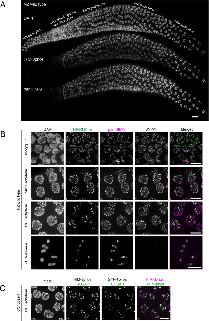
Fig 2. Phosphorylated HIM-3 localizes to the SC and becomes enriched on short arms.
A, Wild-type germline showing DNA stained with DAPI (top), and immunostaining with phosphospecific HIM-3 antibodies (middle), and non-phosphospecific HIM-3 (pan-HIM-3) antibodies (bottom). B, Wild-type N2 oocyte precursor cells taken at different meiotic stages and immunostained for phospho-HIM-3 (green in merged images), SYP-1 and pan-HIM-3 (magenta in merged images). Diakinesis chromosomes (-1 diakinesis nucleus) with cruciform HIM-3 staining are circled in white. Due to the arbitrary orientation of chromosomes relative to the optical axis, cruciform structures of bivalent chromosomes are not always visible. C, Late pachytene cells (gfp::cosa-1) co-immunostained for phospho-HIM-3 (magenta in merged image), phospho-SYP-1 (green in merged image), and anti-GFP showing the position of GFP::COSA-1 (green in two middle images). Scale bars, 5μm.

