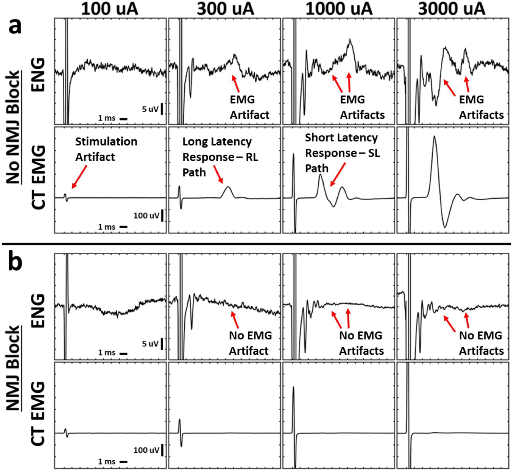
Figure 3.

Stimulation-triggered median ENG and EMG evoked by cervical VNS before and after neuromuscular junction blockade. Electromyograms (EMG) exhibited short- and long-latency components at distinct thresholds, and these signals contaminated the electroneurograms (ENG). Neuromuscular junction blockade with vecuronium eliminated all cricothyroid (CT) and cricoarytenoid (CA, not shown) EMGs and EMG-contamination of ENGs. (a) Simultaneously collected ENG and EMG at multiple stimulation amplitudes (columns) without neuromuscular blockade. (b) Analogous data in the same animal following neuromuscular blockade. X-axis ticks in all plots are 1 ms. Y-axis ticks are 5 μV in all ENG plots and 100 μV in all EMG plots.
