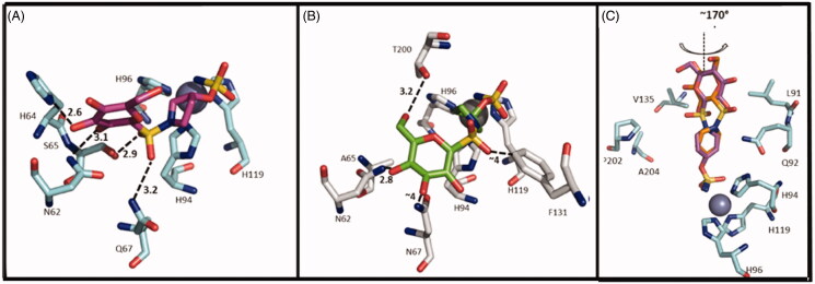
Figure 5.
Interactions at the binding site in the structure of CA IX/57 complex (A), CA II/57 complex (B) and CA IX/55 complex (C) 29. A) CA IX mimic (cyan) and 57 (magenta) (PDB ID: 4R5A). B) CA II (grey) and 57 (green) (PDB ID: 4R59). C) Overlay of the two conformations of 55 (purple and orange) with CA IX mimic. (PDB ID: 4R5B).

