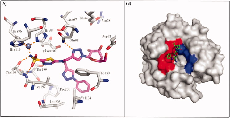
Figure 13.
(A) Interactions at the binding site in the structure of CA II/332(magenta) complex (PDB ID: 4CQ0). (B) Surface representation of CA II/332 complex. The hydrophobic half of CA II is red and the hydrophilic half is blue to highlight the different interactions67.

