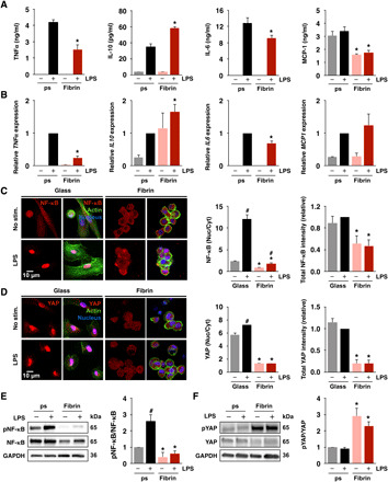
Fig. 1. Culture substrate modulates macrophage inflammatory activation, NF-κB, and YAP.

(A) Cytokine secretion by monocyte-derived macrophages cultured on fibrin gels (2 mg/ml) or polystyrene (ps), stimulated with LPS (10 ng/ml), measured by enzyme-linked immunosorbent assay (ELISA). (B) Relative expression of genes in (A) analyzed by quantitative PCR (qPCR), normalized to polystyrene with LPS. (C) Immunofluorescence confocal images of NF-κB (left) in monocyte-derived macrophages cultured on glass or fibrin (2 mg/ml) for 24 hours and stimulated with LPS for 1 hour and quantification of nuclear/cytoplasmic ratio and total NF-κB intensities (right). (D) Immunofluorescence confocal images of YAP (left) and quantification of nuclear/cytoplasmic ratio and total YAP intensities (right) of the same conditions shown in (C). (E) Immunoblots and quantification of NF-κB and phosphorylated NF-κB (pNF-κB) in monocyte-derived macrophages as in (C). (F) Immunoblots and quantification of YAP and phosphorylated YAP (pYAP; S127) in macrophages cultured as in (C). For immunoblots and immunostaining, quantification is an average of three blots or at least 150 cells across three biological replicates, respectively. All the values are means ± SEM; *P < 0.05 when comparing fibrin to polystyrene or glass with the same soluble condition and #P < 0.05 when comparing LPS to no LPS with the same substrate condition, assessed by two-tailed Student’s t test. GAPDH, glyceraldehyde-3-phosphate dehydrogenase.
