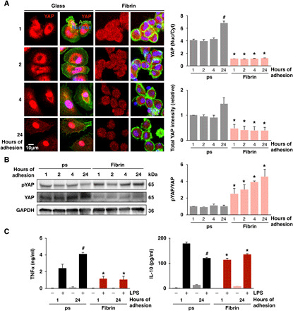
Fig. 2. Adhesion dynamically regulates YAP nuclear translocation and inflammation.

(A) Immunofluorescence confocal images of YAP in monocyte-derived macrophages cultured on glass or fibrin hydrogels (2 mg/ml) for 1, 2, 4, and 24 hours (left) and quantification of nuclear-to-cytoplasmic ratio and total intensity (right). (B) Immunoblot of phosphorylated and total YAP at 1, 2, 4, and 24 hours after adhesion (left) and quantification of phosphorylated to total YAP ratio (right). (C) Secretion of TNFα and IL-10 by monocyte-derived macrophages cultured on polystyrene or fibrin hydrogels (2 mg/ml) for 1 or 24 hours and then stimulated with LPS (10 ng/ml) for 6 hours, analyzed by ELISA. Immunoblots and cytokines are quantified across three separate blots/biological replicates. For immunostaining, analysis was performed on at least 150 cells across three biological replicates at each time point. Data are presented as means ± SEM; *P < 0.05 when comparing fibrin to glass or polystyrene and #P < 0.05 when comparing 1 hour versus 24 hours of adhesion, assessed by two-tailed Student’s t test.
