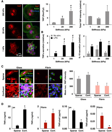
Fig. 3. Substrate stiffness, but not cell confinement, modulates YAP nuclear localization and inflammation.

(A) Immunofluorescence confocal images of YAP in monocyte-derived macrophages cultured on polyacrylamide gels of varying stiffness for 24 hours and stimulated with LPS (10 ng/ml) for 6 hours (left) and quantification of nuclear to cytoplasmic ratio and total intensity (right). (B) Cytokine secretion from cells in (A) analyzed by ELISA. (C) Immunofluorescence confocal images of YAP in monocyte-derived macrophages cultured on glass at low (0.02 million cells/cm2) or high (0.33 million cells/cm2) seeding density (left) and quantification of mean nuclear YAP intensity (right). (D) Secretion of TNFα and IL-10 of cells in (C) and stimulated with LPS (10 ng/ml) for 6 hours, analyzed by ELISA. Immunostaining analysis was performed on at least 150 cells across three biological replicates. Values are means ± SEM of three biological replicates; *P < 0.05 when comparing 1 to 20 kPa or 280 kPa and #P < 0.05 when comparing 20 and 280 kPa in (A), *P < 0.05 comparing 1 to 20 kPa or 280 kPa and #P < 0.05 when comparing LPS to no LPS in (B), and *P < 0.05 when comparing high versus low cell density in (C), assessed by two-tailed Student’s t test.
