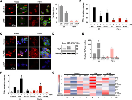
Fig. 4. Nuclear YAP potentiates inflammatory activation of macrophages.

(A) Immunofluorescence confocal images of YAP in monocyte-derived macrophages treated with YAP siRNA (siYAP) or nontarget control (siControl or siC) for 48 hours and cultured on glass or fibrin (left) and quantification of nuclear to cytoplasmic ratio of YAP (right). (B) Secretion of TNFα from macrophages after knockdown of YAP or TAZ and stimulated with LPS (10 ng/ml), analyzed by ELISA. (C) Immunofluorescence confocal images of YAP in YAP-5SA–expressing and shYAP-expressing THP-1 cells or blank lentiviral vector–expressing control cells cultured on glass or fibrin and treated with PMA for 24 hours. GFP, green fluorescent protein. (D) Immunoblot of transgene expression. (E) YAP gene expression analyzed by qPCR of the conditions in (C). (F) Secretion of TNFα from cells in (C) stimulated with LPS (10 ng/ml), analyzed by ELISA. (G) Secretion of proinflammatory cytokines analyzed by multiplex ELISA using human cytokine array for the cells in (C) stimulated with LPS (10 ng/ml). Data are presented as means ± SEM of three biological replicates; *P < 0.05 when comparing siYAP, siTAZ, or YAP-5SA or shYAP versus respective controls, assessed by two-tailed Student’s t test. For multiplex ELISA, data are means of three biological replicates for all the LPS-stimulated conditions. ND, not determined; IFN-γ, interferon-γ; GM-CSF, granulocyte-macrophage colony-stimulating factor.
