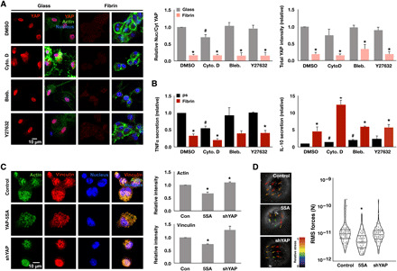
Fig. 5. Actin polymerization is required for YAP, which negatively feeds back to inhibit actin, adhesion, and contractility.

(A) Immunofluorescence confocal images of YAP in monocyte-derived macrophages, cultured on glass or fibrin hydrogel (2 mg/ml) for 24 hours and treated with indicated cytoskeletal drugs for 6 hours (left), and quantification of YAP nuclear-to-cytoplasmic ratio and total intensity (right). (B) Secretion of TNFα and IL-10 secreted from cells in (A) and stimulated with LPS for 6 hours. (C) Immunofluorescence confocal images of actin and vinculin in YAP-5SA–expressing and shYAP-expressing THP-1 cells or blank lentiviral vector–transduced control cells cultured on glass overnight (left) and quantification of total actin and vinculin intensity (right). (D) Representative bead displacement vectors from particle image velocimetry analysis by traction force microscopy (TFM; left) and quantification of root mean square (RMS) forces of at least 60 cells per condition for YAP-5SA–expressing and shYAP-expressing THP-1 cells or blank lentiviral vector–transduced control cells seeded on 10-kPa polyacrylamide gels. Values are means ± SEM across three biological replicates; *P < 0.05 when comparing fibrin versus glass or polystyrene or when comparing YAP-5A and shYAP with control and #P < 0.05 when comparing drug versus dimethyl sulfoxide (DMSO), assessed by two-tailed Student’s t test.
