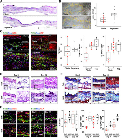
Fig. 6. Biomaterials modulate macrophage YAP and inflammatory activation in vivo.

(A) H&E of fibrin- and Tegaderm-treated wounds, at PWD 5. (B) Micrographs of whole-mount scar tissue at PWD 30 and scar size quantification. (C) Confocal images of iNOS (inflammatory marker), YAP, and F4/80 (macrophage marker) in wound tissue at PWD 5 and quantification of %F4/80+ cells, %F4/80+ YAP+, and %F4/80+ iNOS+. (i to iv) Insets show representative cells in contact or not in contact with fibrin gels, which are labeled as + or – fibrin, respectively, in graphs on the right. (D and E) Representative tissue sections surrounding subcutaneously implanted soft (1 kPa) and stiff (140 kPa) PEGDA hydrogels for 3 or 14 days, stained with H&E (D) or Masson’s trichrome (E). (F) Confocal images of immunohistochemistry and quantification as in (C), for soft and stiff implants after 3 and 14 days. The asterisks (*) indicate hydrogel material in (D to F). n ≥ 5 animals and *P < 0.05 when comparing fibrin contact versus no contact or Tegaderm (C) or soft versus stiff (F), assessed by two-tailed Student’s t test. Photo credit: Raji Nagalla, University of California, Irvine.
