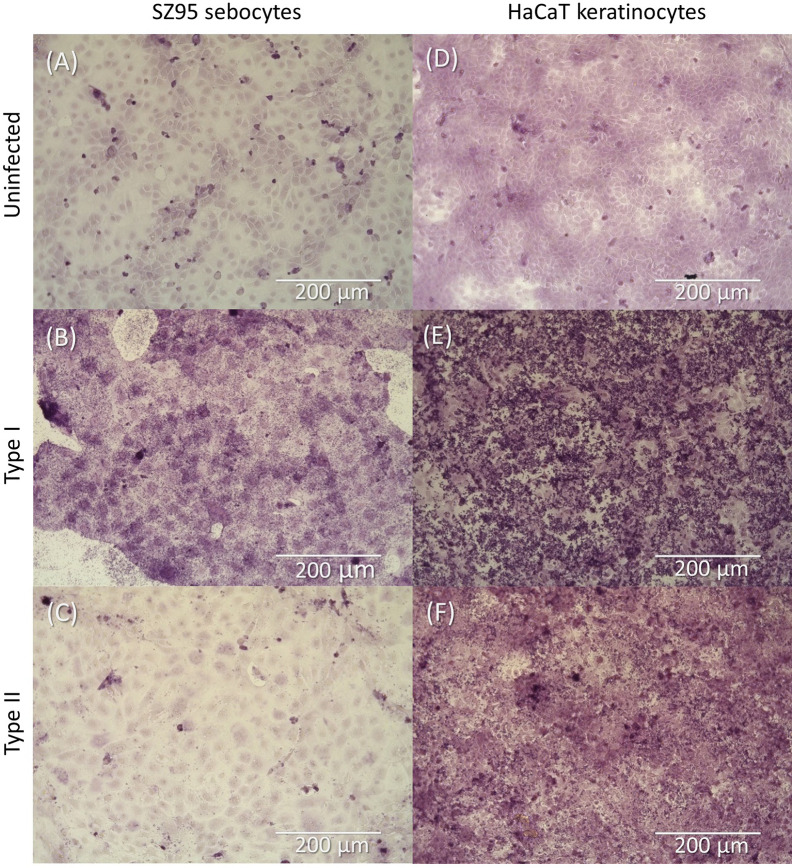
Figure 2.
Light microscopy image of SZ95 sebocytes (A–C) and HaCaT keratinocytes (D–F) after infection with C. acnes and a modified Gram stain. (A) Uninfected SZ95 cells, (B) SZ95 cells infected with HL053PA1 (type I strain), and (C) SZ95 cells infected with HL110PA3 (type II strain). (D) Uninfected HaCaT cells, (E) HaCaT cells infected with HL053PA1, and (F) HaCaT cells infected with HL110PA3. Total magnification: 368x. Scale bars: 200 µm.

