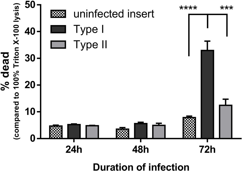
Figure 8.
HaCaT cell viability during infection with type I or type II C. acnes strains was monitored using LDH activity assays. Cell viability is expressed as the percentage of dead cells compared to 100% dead cells after lysis with 1% Triton X-100 in PBS. Data shown are the mean from at least three biological replicates, error bars indicate SEM. ***p < 0.005, ****p < 0.001.

