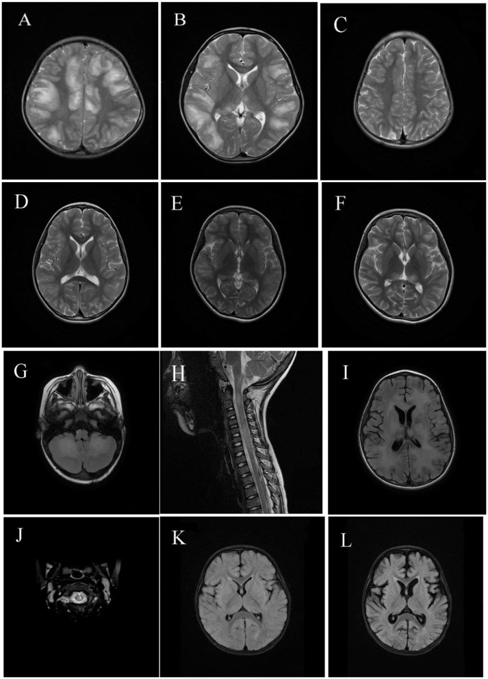
Figure 2.
Magnetic resonance imaging (MRI) results of pediatric acute disseminating encephalomyelitis (ADEM) with myelin oligodendrocyte glycoprotein antibodies (MOG-abs). (A–D) Cerebral MRI of a 6-year-old patient with MOG-abs, who presented with fever, headache, projectile vomiting, drowsiness, and ataxia, revealed large and blurred lesions involving both hemispheres [(A); axial-T2)], corpus callosum, thalamus/basal ganglia [(B); axial-T2], brainstem, and cerebellum (not shown). Three months later, supratentorial and thalamus/basal ganglia lesions had mostly resolved [(C,D); axial-T2]. (E,F) Cerebral MRI of a 10-year-old patient with anti-MOG-abs, who presented with fever, headache, extreme fatigue, and lethargy, revealed small and blurred lesions involving both hemispheres and the thalamus/basal ganglia [(E); axial-T2]; 10 days later, lesions in both hemispheres and the thalamus/basal ganglia enlarged even after immunotherapy [(F); axial-T2]. (G–I) Cerebral and spinal MRI of a 5-year-old patient with anti-MOG-abs, who presented with slight fever, somnolence, and abnormal behavior, showed large and blurred cerebellar and brainstem lesions [(G); axial-T2], LETM [(H); sagittal-T2], supratentorial white matter lesions [(I); axial-T2], and thalamus/basal ganglia (not shown). (J) Spinal MRI of a 5-year-old patient with anti-MOG-abs, who presented with slight fever, drowsiness, dysarthria, and ataxia, revealed small and blurred lesions of the cervical spinal cord [(J); axial-T2] and large and blurred lesions of the basal ganglia, brainstem, and supratentorial lesions (not shown). [(K,L); axial-T2] Cerebral MRI of a 1-year-old patient with anti-MOG-abs, who presented with slight fever and had coma and was admitted in the intensive care unit, revealed small, blurred supratentorial and brainstem lesions (not shown); 3 weeks later, a repeat cerebral MRI revealed cortical atrophy.

