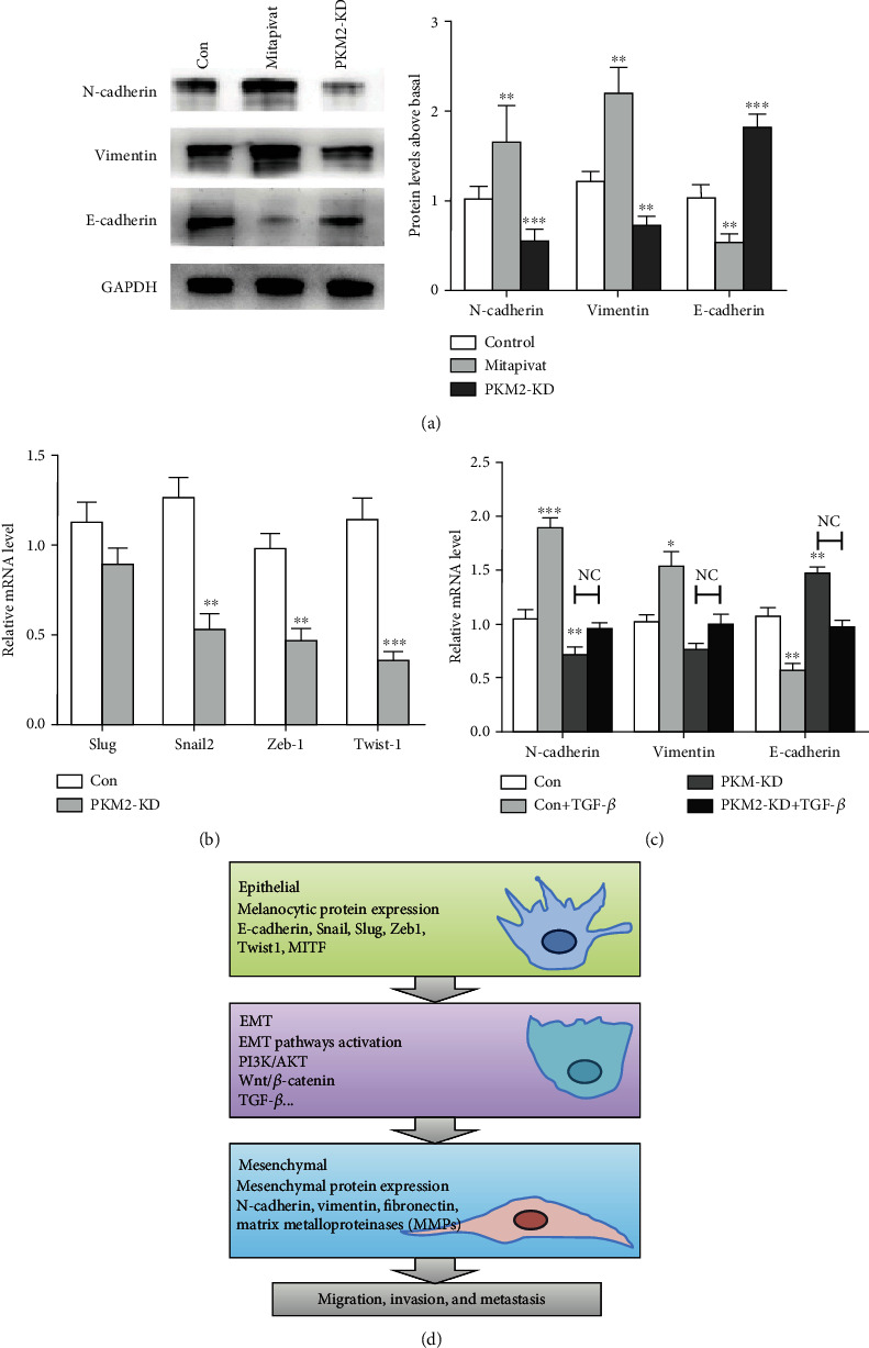
Figure 5.

Silencing PKM2 suppresses MDA-MB-231 cell EMT. (a) Western blot analysis of protein levels of EMT marker proteins in control, Mitapivat, and PKM2-KD groups. GAPDH was used to normalize protein expression (left panel). The protein levels of EMT markers were quantified and compared (right panel). (b) Q-RT PCR analysis of mRNA levels of key EMT-related genes (Snail, Slug, Zeb1, and Twist1) in control and PKM2-KD cells normalized to GAPDH. (c) Relative mRNA levels of vimentin, N-cadherin, and E-cadherin after EMT induction in MDA-MB-231 cells in control and PKM2-KD cells for 48 h. (d) Changes involved in EMT (∗∗∗P < 0.001, ∗∗P < 0.01, NC : P > 0.01).
