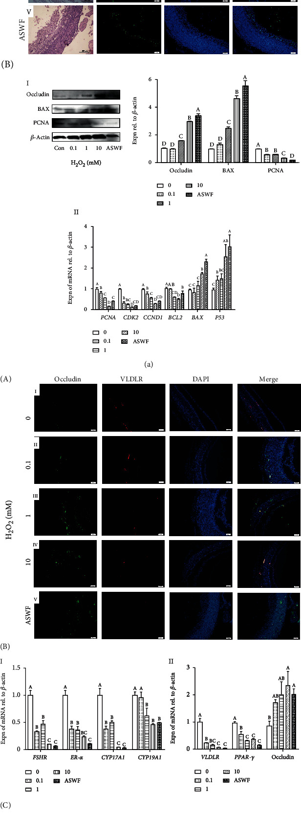
Figure 4.

(a) Effect of H2O2 on cell apoptosis in the atretic follicles. A: H&E staining and TUNEL assay in the SWFs (treated with H2O2) and ASWFs. Scale bar: 100 μm for H&E staining and 50 μm for TUNEL assay. B-I: Western blot and gray analysis of occludin, BAX, and PCNA expression after H2O2 treatment. B-II: the expression of PCNA, CDK2, CCND1, BCL2, BAX, and P53 mRNAs in the SWFs (treated with H2O2) and ASWFs. (b) Effect of H2O2 on yolk deposition capacity and steroidogenesis in the atretic follicle model by H2O2. A: immunofluorescent labels with occludin (green), VLDLR (red), and DAPI (blue) in the histological sections of the SWFs with different concentrations of H2O2. Scale bar: 50 μm. B-I: the expression of FSHR, ER-α, CYP17A1, and CYP19A1 mRNAs. B-II: the expression of yolk deposition-related genes VLDLR, PPAR-γ, and occludin. C: Western blot and grey analysis of ER-α and FSHR after H2O2 treatment. Values represent the means ± SEM of three replicates in each group. Different lowercase letters indicate significant difference (P < 0.05).
