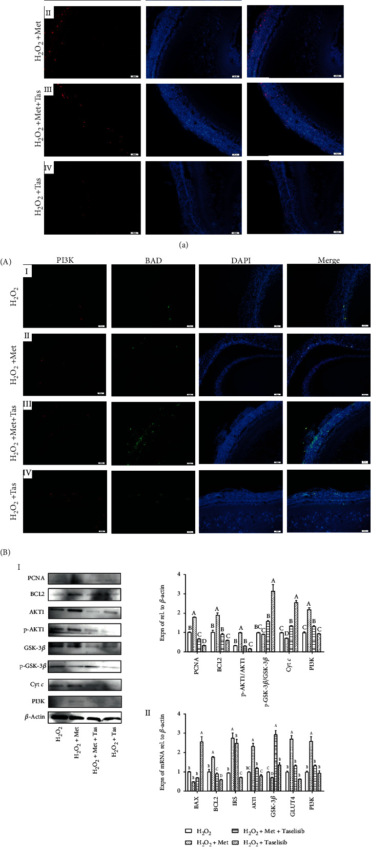
Figure 8.

(a) Effect of Met and Taselisib (Tas) on BrdU incorporation in proliferating cells in the H2O2-induced ASWFs. A: red—BrdU-labelled cells; blue—DAPI staining. Scale bar: 50 μm. (b) Function of PI3K in Met is to stimulate cell proliferation in GCs of ASWFs. A: effect of Met and Tas on expression of PI3K (red) and BAD (green) in ASWFs. Blue: DAPI staining. Scale bar: 50 μm. B-I: Western blot and grey analysis of PCNA, BCL2, p-AKT1, AKT1, p-GSK-3β, GSK-3β, Cyt c, and PI3K expression in ASWFs. B-II: qRT-PCR analysis of the BAX, BCL2, IRS, AKT1, GSK-3β, GLUT4, and PI3K mRNA expression in ASWFs. Values represent the means ± SEM of three replicates in each group. Different lowercase letters indicate significant difference (P < 0.05).
