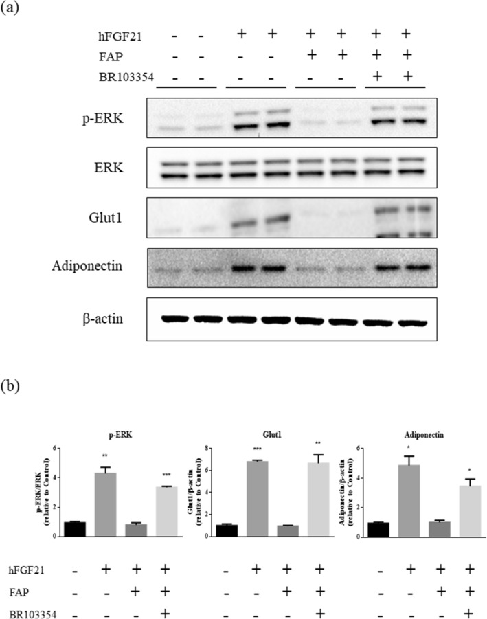
Figure 2.
Enhanced ERK signaling in 3T3/L1 cells by BR103354. (a) Fully differentiated 3T3/L1 adipocytes were incubated with full-length hFGF21 at 500 ng/mL. FGF21 was pre-incubated with PBS or recombinant FAP at 100 ng/mL (substrate: enzyme ratio = 5:1) for 2 h, with or without 5 µM of BR103354. Phosphorylated ERK (p-ERK), total ERK (ERK), Glut1, adiponectin and β-actin were measured by immunoblots. (b) Quantification of p-ERK, Glut1 and adiponectin was obtained with densitometric analysis, and normalized with total ERK or β-actin, respectively. All data are the means ± S.D. *P < 0.05, **P < 0.01 and ***P < 0.001 vs. no treatment (control) group.

