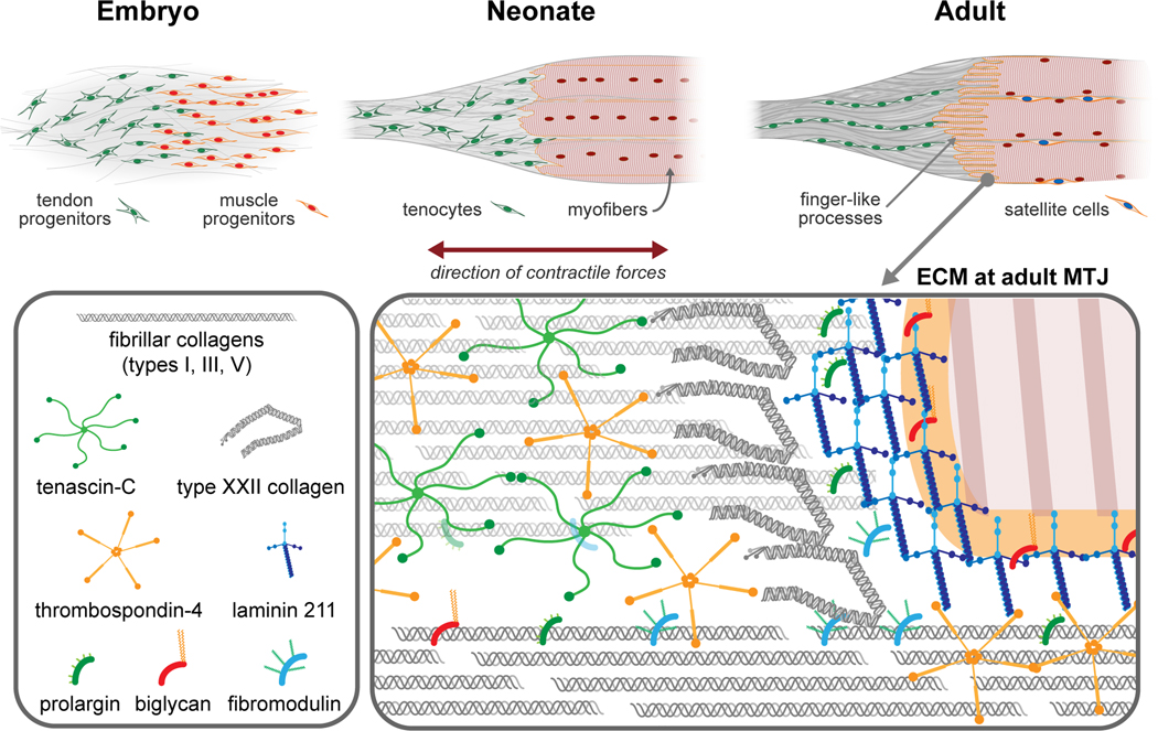
Figure 1:
Development of the MTJ (top). During embryogenesis, tendon and muscle progenitors condense at the site of future MTJ. In the neonate, the muscle - tendon interface appears smooth and there are very few finger-like processes present. In the adult, muscle fibers are mature and perfectly aligned at the MTJ. Finger-like extensions of the myofibers are well established and integrated with the tendon structure. The tendon ECM and cells are aligned along the direction of the load. Distribution of ECM discussed in this review within MTJ (bottom).

