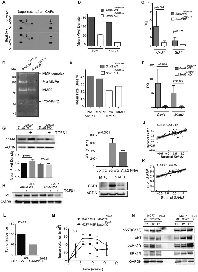
Fig. 6. SNAI2 in the stroma modified the microenvironment required to support cancer behavior.

(A) Mouse CAFs (mCAFs) obtained from tumors developed in Snai2 WTErbB2++, and Snai2 KOErbB2++ parous mice were maintained for three days in the absence of serum. The supernatant was analyzed using a mouse cytokine array. Arrows point to differences in CXCL-1 (left) and SDF-1(right) protein expression. (B) Quantification of the cytokine array showed in panel A with the ImageJ software. (C) Relative Cxcl1 and Sdf1 mRNA expression in sorted CD140+/CD31−/EpCAM− cells from tumors developed in WT (N=8) or KO (N=9) parous mice, determined by real-time QPCR (Mann-Whitney U test). (D) Gelatin zymography assay on the supernatant from cultured mCAFs. Supernatants were collected and separated by electrophoresis, and gelatinase activity was visualized by standard staining. (E) Gelatin zymography bands were quantified using the ImageJ software. (F) Relative Cxcl1 and Mmp2 mRNA expression in sorted CD140+/CD31−/EpCAM− cells from metastatic tumors developed in WT and KO parous mice, as determined by real-time QPCR (Mann-Whitney U test). (G) Primary WT or KO mouse embryonic fibroblasts (MEFs) were treated with TGFβ1 (10 ng/mL) for 48 hours in the absence of serum, and αSMA (Student's t-test, N=3), or FAP (H) expression was determined by western blots. (I) Relative SDF1 mRNA expression in WT or SNAI2 depleted human CAFs (hCAFs) determined by real-time QPCR (one-way ANOVA, upper graph) and the SDF1 protein levels were assessed by western blot (lower image). (J, K) A positive correlation between the levels of SNAI2 and (J) SDF1 or (K) FAP was found in the stroma of breast tumors (Pearson's correlation). (L) Incidence of tumors generated by xenografts of WT or Snai2-deficient MEFs mixed with MCF7 cells (Chi-squared test, N=6). (M) Volume of tumors generated by MCF7 cells co-injected with WT or Snai2-deficient primary MEFs in subcutaneous xenografts (tumor volumes were compared using t-tests, N=6). (N) The tumors shown in panel M were analyzed for total and phosphorylated ERK and AKT levels by western blots (N=3).
