
Extended Data Fig. 3.
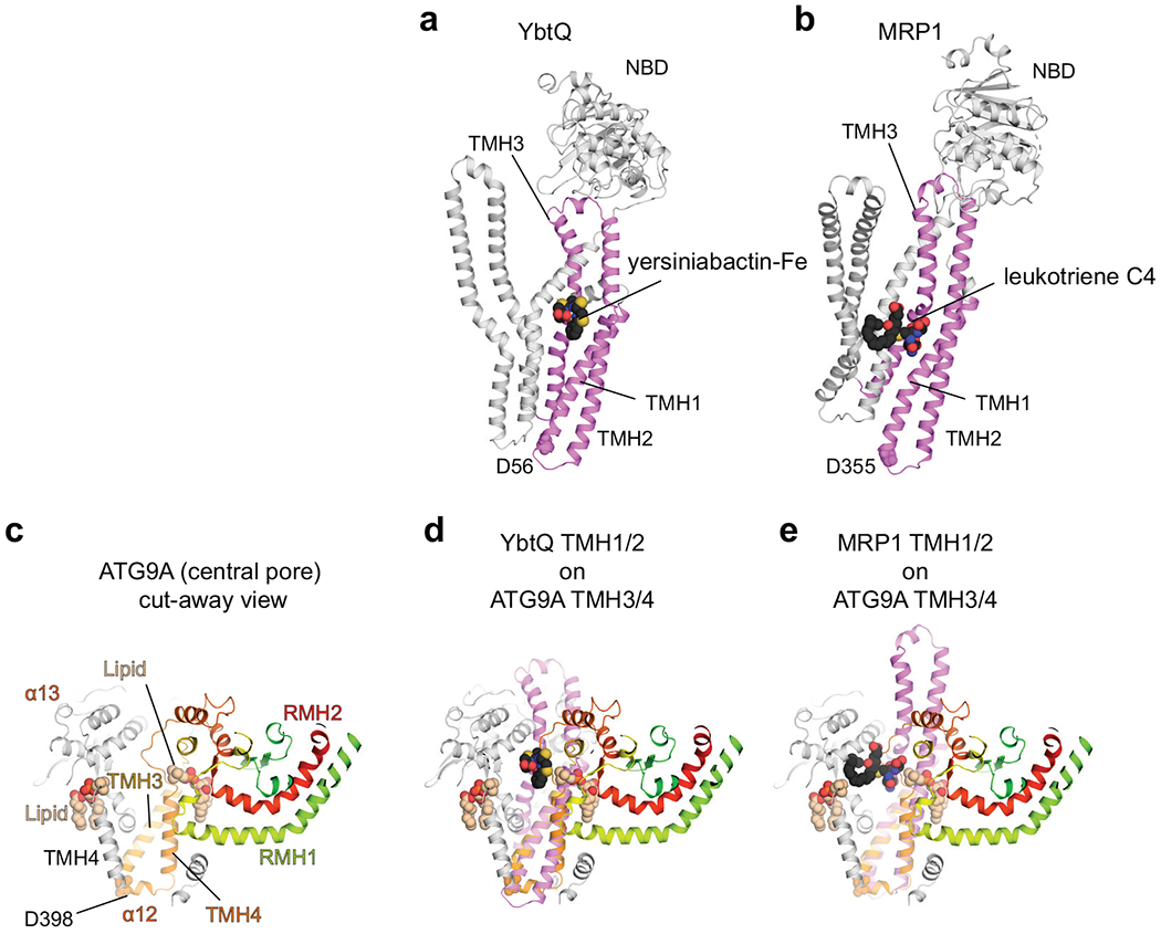
Structural alignments of ATG9A with ABCexps. a,b, Structures of YbtQ (a, PDB 6P6J) and MRP1 (b, PDB 5UJA) bound to their substrates. The TMH1–3s are colored violet. c, A cut-away view of the central pore of ATG9A. d,e, Superpositions of ATG9A with YbtQ (d) and MRP1 (e). ATG9A’s TMH3/TMH4 can be superimposed with TMH1/TMH2 from the identified ABCexp structures using as a guide for the helix register the conserved Asp (D398 in ATG9 with D53 in MsbA, D56 in YbtQ, D355 in MRP1). In all cases, ABCexps’ substrates locate to the central pore of ATG9A.