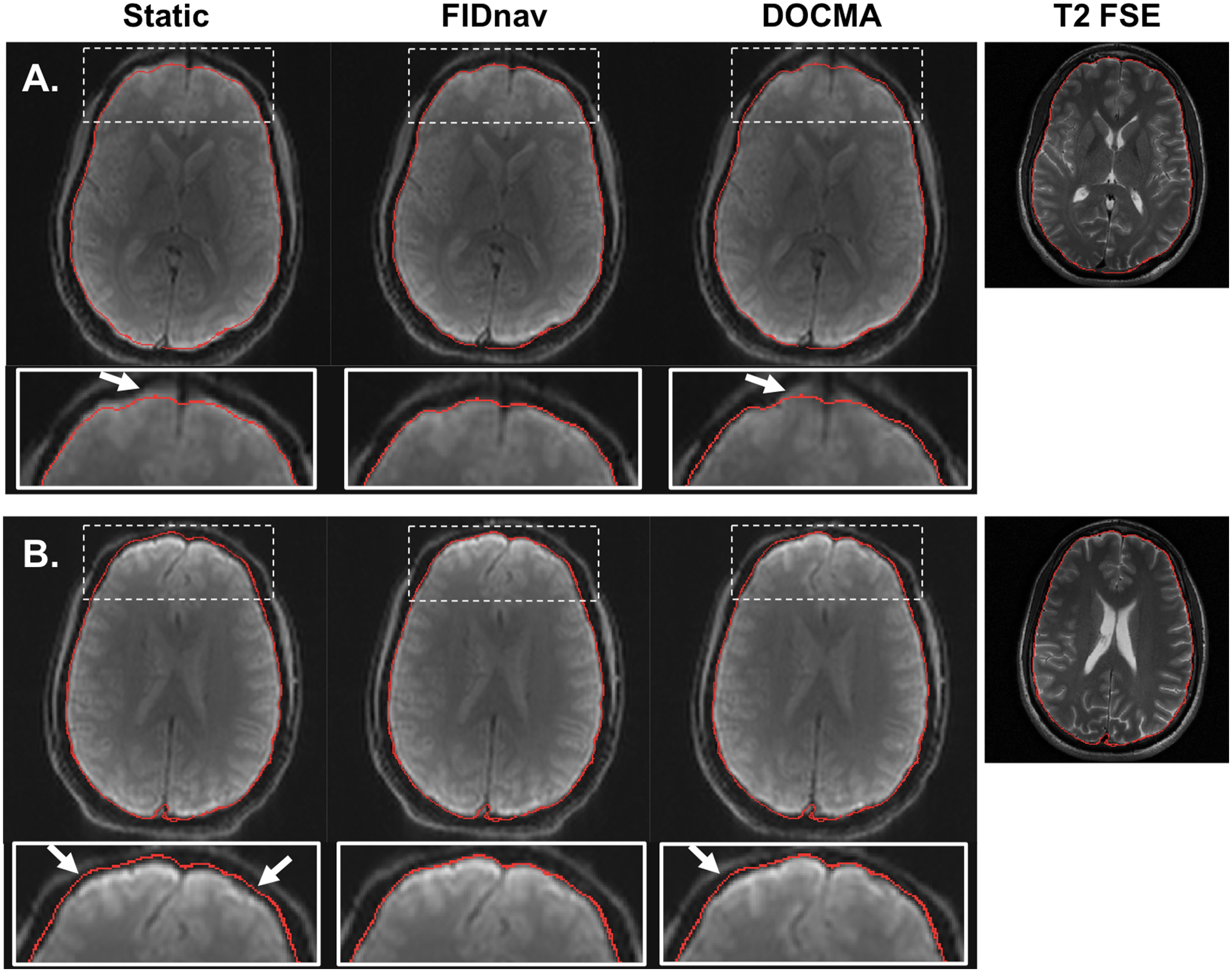
Figure 5.

Distortion correction results in a representative volunteer following correction with a static voxel shift map (VSM) and with VSMs obtained from FIDnav and DOCMA field measurements for changing Y-shim (A) and nose touching (B). The brain boundary extracted from T2 FSE (shown on the right for comparison) is overlaid on each corrected image. Arrows denote regions with residual unwarping errors, observed with both static and DOCMA correction. FIDnavs enable reliable geometric distortion correction for both shim changes and nose touching.
